|
|
||
14/07/10 |
|
|
Aim: Transformation of Fusarium graminearum for introduction of a deletion vector construct. (for construction of the used vectors please see the following page...)
Day 1: 10 ml of LB medium (pH > 7.7) supplemented with 50 µg/ml kanamycin (and 10 µg/ml rifampicin) in a 100 ml Erlenmeyer flask is inoculated with A. tumefaciens LBA4404 pAg1-H3::Dgene and incubate for one to two days at 28 oC with shake at 100 rpm.
Day 2: 1. Place 11 black AGF220 80 mm filters in a glass Petri dish and autoclave it. 2. Prepare 50 ml liquid IMAS medium. 3. Inoculate 10 ml IMAS + kanamycin (50 µg/ml) in 100 ml Erlenmeyer flasks with 100 µl, 200 µl and 300 µl LBA4404-Dgene and incubate at 28 oC with shake (100 rpm) until OD600 reaches 0.5 – 0.7 (typically the next day) in one of the cultures.
Day 3: 1. Cast 13 IMAS plates (10 for transformation and 3 for controls). Label plates with “IMAS - Dgene #1 to #10” date and your initials. The plates can also be made on day 2, just remember to store them at 5 oC. 2. Place one sterile AGF220 80 mm filter onto the 11 of the 13 IMAS plates. Make sure that no air pockets exist between the medium and filters, this is done by adding 50 to 400 µl of sterile water (depending on the moisture of the plates) onto the centre of the filters and spreading it with a sterile Drigalsky spatula. 3. When the LBA4404 cells has reached an OD600 of 0.5– 0.7, dilute the F. graminearum PH1 (Hyg(s)) spores with liquid IMAS medium to a final spore concentration of 2*106 spores/ml (a total of 1.2 ml is needed for the following steps = 2.4*106 spores). 4. Mix the LBA4404 culture in a 1:1 (v:v) ratio with the F. graminearum PH1 (Hyg(S)) spores (resulting in 1*106 spores/ml). 5. Spread 200 ml of the LBA4404/F. graminearum spore solution onto each of the sterile filters (equalling 2*105 spores/plate) using a sterile Drigalsky spatula. Make the following control plates: 1. Sterile filters on IMAS-plates 2. A. tumefaciens Dgene 100 µl alone (without filter) 3. F. graminearum PH1 (Hyg(S)) alone (2*105 spores) (without filter) 6. Incubate the 13 plates for 2-3 days at 25 - 28 oC in darkness.
Day 4: 1. Cast 10 DFM plates supplemented with 150 µg/ml hygromycin and 300 µg/ml mefoxin. Label: “S1 - Dgene #X” (S1 = 1st selection round).
Day 6: 1. Under sterile conditions transfer the filters onto the DFM + hygromycin + mefoxin plates. Discard the IMAS plates. 2. Incubate the 10 plates at 25 oC for 3-5 days.
Day 10 1. Cast 10 new DFM plates supplemented with 150 µg/ml hygromycin Label: “S2 - gene #X” (S2 = 2nd selection round).
Day 11: 1. Under sterile conditions transfer the filters onto the DFM + hygromycin plates. Store the DFM plates from 1. selection round at room temperature in airtight bags. 2. Incubate the 10 S2 plates at 25 oC for 3 to 5 days, depending on the growth rate of the transformants.
Day 13: 1. Cast 5.5 cm DFM plates supplemented with 100 µg/ml hygromycin, the number depending on the number of transformants. Label: Fg Dgene #T1 (T = transformant)
Day 14: 1. When colonies have spread into the medium, the filters are discarded and visible colonies are transferred to 5.5 cm DFM plates + 100 µg/ml hygromycin as they appear. Isolate both transformants with a wild type phenotype and new phenotypes. The isolation is best done by sticking a sterile toothpick into the colony (through the agar) and then repeating the action on the centre of the isolation plate. 2. Incubate the plates for 2 to 7 days.
Day ?: 1. Screen the isolated transformants by fungal colony-PCR (see other protocol) with the following primers: a. Located in the deleted region (primers gene-T1/T2) b. Located in HygB and outside the right recombination flank (gene-T3) c. Located in HygB and outside the left recombination flank (gene-T4)
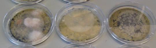
Examples of S2 plates with filters:
Note that some plates have very visible fluffy colonies on top of the filters, while others seems to be empty. However, many times the "empty" plates also yield correct transformants, so do not discard them, they just need to incubate a little longer. Examples of S2 plates without filters:
Note the transformants have grown from the filters and into the medium.
|
Dette sted blev sidst opdateret 14. July 2010